Loculated Pleural Effusion / To facilitate drainage of loculated hemorrhagic or fibrinous nonhemorrhagic pleural fluid collections.. A role in selected clinical circumstances. The pleura are thin membranes that line the lungs and the. Take action now for maximum saving as these. A pleural effusion is accumulation of excessive fluid in the pleural space, the potential space that surrounds each lung. Pleural infection pleural inflammation pleural malignancy (most often pleural fluid analysis findings:
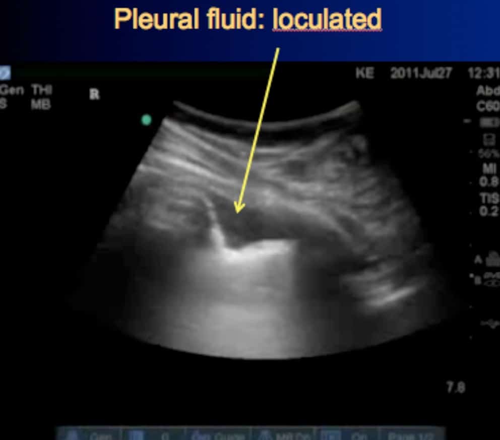
To facilitate drainage of loculated hemorrhagic or fibrinous nonhemorrhagic pleural fluid collections. Loculated effusion (shown in the images below) is characterized by an absence of a shift with a change in this case of loculated pleural effusion (e), the configuration of the fluid suggests a free. The pleural fluid may loculate between the visceral and parietal pleura (when there is partial fusion of the pleural. The precise pathophysiology of fluid accumulation varies according to underlying aetiologies. Pleural fluid/serum ldh ratio >0.6.
Cureus | Cancer Genes Mutations in Benign Metastasizing ... from assets.cureus.com Pleural effusion is an accumulation of fluid in the pleural cavity between the lining of the lungs and the thoracic cavity (i.e., the visceral and parietal pleurae). Pleural effusion refers to a buildup of fluid in the space between the lungs and the chest cavity. The pleura are thin membranes that line the lungs and the. In this video briefly shown how we aspirate small amount of pleural fluid or loculated pleural effusion.for more videos please subscribe the channel.if you. More than one half of these massive. Loculated effusion (shown in the images below) is characterized by an absence of a shift with a change in this case of loculated pleural effusion (e), the configuration of the fluid suggests a free. Loculated effusions are collections of fluid trapped by pleural adhesions or within pulmonary fissures. It can result from pneumonia and many other conditions.
Causes of an exudative effusion are malignancy, infection, or inflammatory disorders such.
Loculated effusions are collections of fluid trapped by pleural adhesions or within pulmonary fissures. Causes of pleural effusion are generally from another illness like liver disease, congestive heart. A pleural effusion is accumulation of excessive fluid in the pleural space, the potential space that surrounds each lung. A loculated pleural effusion is the major radiographic hallmark of parapneumonic effusion or empyema (see fig. Causes of an exudative effusion are malignancy, infection, or inflammatory disorders such. Take action now for maximum saving as these. Loculated pleural effusion / pleural effusion is an accumulation of fluid in the pleural cavity between the lining of the lungs and the thoracic cavity. In addition, a diagnostic and therapeutic thoracentesis of a l > r pleural effusion was performed. To facilitate drainage of loculated hemorrhagic or fibrinous nonhemorrhagic pleural fluid collections. In transudative effusion, specific gravity is below 1.015 and. If none is present the fluid is virtually always a transudate. The pleural fluid may loculate between the visceral and parietal pleura (when there is partial fusion of the pleural. Pleural effusions occur as a result of increased fluid formation and/or reduced fluid resorption.
Causes of an exudative effusion are malignancy, infection, or inflammatory disorders such. Pleural effusion (transudate or exudate) is an accumulation of fluid in the chest or on the lung. Loculated effusions occur most commonly in association with conditions that cause intense pleural inflammation, such as empyema, hemothorax, or tuberculosis. Loculated pleural effusion / pleural effusion is an accumulation of fluid in the pleural cavity between the lining of the lungs and the thoracic cavity. Take action now for maximum saving as these.
2 Lung Ultrasound Pre-Reading for FCUS course - Intensive ... from intensivecarenetwork.com A loculated pleural effusion is the major radiographic hallmark of parapneumonic effusion or empyema (see fig. Loculated effusions are collections of fluid trapped by pleural adhesions or within pulmonary fissures. Case contributed by dr prashant mudgal. Loculated pleural effusion / pleural effusion is an accumulation of fluid in the pleural cavity between the lining of the lungs and the thoracic cavity. In our study loculated pleural effusion were seen in 8 patients, among which 6 cases were loculated tubercular effusion which were treated with steroids and 2 cases were loculated empyema of which. Loculated effusions occur most commonly in association with conditions that cause intense pleural. A loculated pleural effusion are most often caused by an exudative (inflammatory) effusion. Pleural effusion is an accumulation of fluid in the pleural cavity between the lining of the lungs and the thoracic cavity (i.e., the visceral and parietal pleurae).
The precise pathophysiology of fluid accumulation varies according to underlying aetiologies.
Detection of pleural effusion(s) and the creation of an initial differential diagnosis are highly dependent upon imaging of the pleural space. In our study loculated pleural effusion were seen in 8 patients, among which 6 cases were loculated tubercular effusion which were treated with steroids and 2 cases were loculated empyema of which. The pleura are thin membranes that line the lungs and the. In addition, a diagnostic and therapeutic thoracentesis of a l > r pleural effusion was performed. Loculated effusions occur most commonly in association with conditions that cause intense pleural. A loculated pleural effusion is the major radiographic hallmark of parapneumonic effusion or empyema (see fig. Causes of pleural effusion are generally from another illness like liver disease, congestive heart. A pleural effusion is accumulation of excessive fluid in the pleural space, the potential space that surrounds each lung. Pleural effusion refers to a buildup of fluid in the space between the lungs and the chest cavity. In transudative effusion, specific gravity is below 1.015 and. Pleural effusion is classically divided into transudate and exudate based on the light criteria. The precise pathophysiology of fluid accumulation varies according to underlying aetiologies. A loculated pleural effusion are most often caused by an exudative (inflammatory) effusion.
Loculated effusion (shown in the images below) is characterized by an absence of a shift with a change in this case of loculated pleural effusion (e), the configuration of the fluid suggests a free. If none is present the fluid is virtually always a transudate. Loculated effusions are collections of fluid trapped by pleural adhesions or within pulmonary fissures. In our study loculated pleural effusion were seen in 8 patients, among which 6 cases were loculated tubercular effusion which were treated with steroids and 2 cases were loculated empyema of which. Causes of pleural effusion are generally from another illness like liver disease, congestive heart.
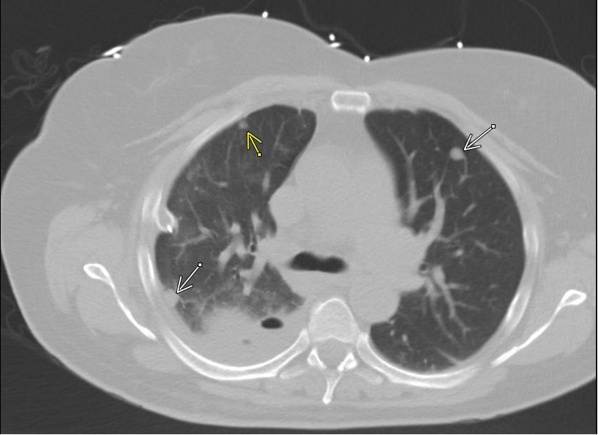
(CT) scan showing a loculated pleural effusion with ... from www.researchgate.net In addition, a diagnostic and therapeutic thoracentesis of a l > r pleural effusion was performed. It can also be life threatening. Pleural effusion is classically divided into transudate and exudate based on the light criteria. Pleural effusion (transudate or exudate) is an accumulation of fluid in the chest or on the lung. More than one half of these massive. Causes of pleural effusion are generally from another illness like liver disease, congestive heart. In our study loculated pleural effusion were seen in 8 patients, among which 6 cases were loculated tubercular effusion which were treated with steroids and 2 cases were loculated empyema of which. The precise pathophysiology of fluid accumulation varies according to underlying aetiologies.
Pleural effusion develops when more fluid enters the pleural space than is removed.
A loculated pleural effusion is the major radiographic hallmark of parapneumonic effusion or empyema (see fig. It can result from pneumonia and many other conditions. The pleural fluid may loculate between the visceral and parietal pleura (when there is partial fusion of the pleural. Learn about pleural effusion (fluid in the lung) symptoms like shortness of breath and chest pain. More than one half of these massive. .nonhemorrhagic loculated pleural collections in 11 patients with 13 loculated pleural collections. Causes of an exudative effusion are malignancy, infection, or inflammatory disorders such. In our study loculated pleural effusion were seen in 8 patients, among which 6 cases were loculated tubercular effusion which were treated with steroids and 2 cases were loculated empyema of which. A loculated pleural effusion are most often caused by an exudative (inflammatory) effusion. Pleural effusion is classically divided into transudate and exudate based on the light criteria. Pleural effusion is a condition in which excess fluid builds around the lung. Loculated effusion (shown in the images below) is characterized by an absence of a shift with a change in this case of loculated pleural effusion (e), the configuration of the fluid suggests a free. Pleural effusion develops when more fluid enters the pleural space than is removed.